Modern methods for tadpole studies
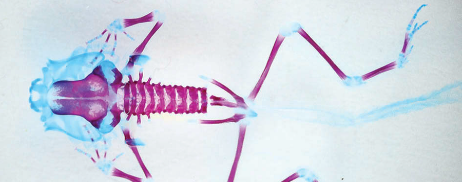
Micro-computed tomography (micro-CT) scanning and three-dimensional reconstruction have revolutionized morphological studies. Whereas species descriptions and comparative studies formerly were based on external appearance and dissection, we now can visualize muscles, skeleton and viscera in intact animals. In most cases, visualization of internal structures depends on appropriate staining methods. Staining with iodine, phosphotungstic acid (PTA) and osmium tetroxide are established methods, but some problems remain. Agents like osmium tetroxide are toxic and the contrast of cartilage generally is unsatisfactory with osmium tetroxide, iodine or PTA. Furthermore, staining results vary for different animals and different developmental stages. We investigated critical point drying as an inexpensive, nontoxic and rapid alternative to staining for frog tadpoles. Critical point drying enables visualization of cartilage and its differentiation from muscle tissue. Shrinkage generally is acceptable. We also present a protocol for clearing and staining frog tadpoles.


