Scanning electron microscopy lab
What is scanning electron microscopy (SEM)?
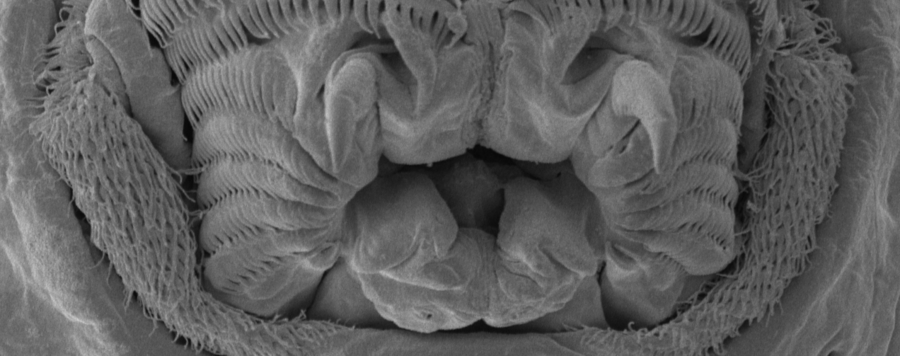
In scanning electron microscopy an electron beam scans the surface of a sample. The interactions between the electrons and the object create an image. In contrast to light microscopy, SEM images have a much higher depth of field and allow stronger magnifications: while light microscopies reach approximately magnifications of 2 000x, SEMs can reach up to 1 000 000x.
In a standard SEM, the electron beam is created in a high vacuum. Thus the studied sample has to be vacuum-tolerant, i.e. it has to be free of water which is usually achieved by critical-point drying. Another problem are electric charges, which are created during the study of nonconductive samples. To avoid these charges, the studied specimen is covered with a very thin layer of gold or another noble metal (sputter coating).
Which equipment is available at ZFMK?
The ZFMK has a Zeiss Gemini Sigma 300 VP SEM. This machine is an environmental scanning electron microscope (ESEM), which means that only the area where the electron beam is created is under high vacuum, while the sample chamber and the detector are in a lower vacuum. As a result, samples can be studied without water-removal or sputter coating which is especially benefiting for museum material.
What is the SEM used for at ZFMK?
It is mainly used for documentation and descriptive purposes. In contrast to light microscopy, much higher magnifications can be achieved what allows for example the study of ultrastructure.


